Today, when precision medicine and minimally invasive diagnosis and treatment have become mainstream, endoscopes are the "eyes" for doctors to explore the mysteries of the human body. Behind these "eyes", the image signal processor (ISP) plays the role of the "visual cortex". Its performance directly defines the quality of the images seen by the doctor and is a crucial link in the modern medical diagnosis chain.
As the core "brain" of the endoscope system, ISP is responsible for processing the original, rough optical signals captured by the front-end image sensor into clear, true-color, and richly detailed medical images in real time. With the rapid evolution of technology, especially the rapid maturity of the domestic medical equipment industry chain, endoscope ISP technology is undergoing profound changes from algorithms to hardware platforms, and is showing a clear localization development trend.
Chapter 1: The Uniqueness of Medical ISP: Why is it Different?
Although image signal processor (ISP) technology has been highly popular in consumer electronics fields such as smartphones and security monitoring, when it is placed in the context of medical endoscopy, its connotation, mission and technical requirements have all undergone fundamental changes. If the consumer ISP is a "makeup artist" who pursues visual beauty, and its goal is to create images that are pleasing and even transcend reality; then the medical ISP is a rigorous "forensic scientist" whose only responsibility is to infinitely approach and objectively present the "absolute reality", because this "reality" is directly related to the accuracy of diagnosis and the life safety of the patient.
This huge difference in roles determines that there are four deep gaps between the endoscope ISP and the general ISP in terms of design philosophy and technical implementation.
1. From “subjective beauty” to “objective reality”: the huge difference in image fidelity
Generic ISPs are designed to please the human eye. It will algorithmically render bluer skies, brighter flowers, and smoother portrait skin. This processing is essentially a kind of "beautification" that allows a certain degree of distortion in exchange for a more attractive look and feel. Medical ISP strictly adheres to the principle of "what you see is what you get", and its requirements for image fidelity have reached a scientific level.
- Absolute color accuracy: In clinical diagnosis, subtle differences in tissue color are key clues to determine pathological status. For example, mild redness of the mucosa may indicate early inflammation, whereas abnormal pallor or dullness may point to ischemia or necrosis. Medical ISP must establish a "medical grade" color calibration system to ensure that the red of blood, the pink of mucous membranes, and the yellow of bile can be accurately restored under complex intracavity light reflection. Any color “optimization” without clinical validation is not allowed as it may mask or falsify pathological features and thereby mislead doctors in their diagnosis.
- Complete preservation of details: Consumer-grade ISPs may smooth out tiny noise or irregular textures in images as "imperfections." But in endoscopic images, these "flaws" may be the rough surface of early cancerous tissue, the outline of tiny polyps, or abnormal blood vessel texture. The medical ISP algorithm must protect these detailed information related to diagnosis to the greatest extent while effectively reducing noise, ensuring that every pixel of the image carries valid medical data.
2. From “acceptable delay” to “zero tolerance delay”: the speed of life and death in real-time
When it comes to taking photos or making video calls on your phone, a delay of a few tenths of a second may only be a minor inconvenience. But in medical procedures, especially surgeries, there is "zero tolerance" for delays. Imagine performing a delicate vascular dissection laparoscopically, with every movement measured by the doctor in millimeters. If the image displayed on the screen lags behind the doctor's hand operation by even 100 milliseconds, this delay may cause the instrument to accidentally damage nerves or blood vessels, causing irreparable consequences. Therefore, medical ISP must have extraordinary processing efficiency to ensure that the end-to-end delay of the entire process from the image sensor to the picture, through the complex processing of the ISP, and finally displayed on the screen is strictly controlled within 150 milliseconds, and even lower requirements are required for surgical-grade applications. This is a kind of "life and death speed", which is the ultimate test of the ISP's computing power and pipeline architecture.
3. From "general algorithm" to "special algorithm": in-depth customization for diagnosis
The algorithm libraries of general ISPs are usually standardized, such as scene recognition, face beautification, night scene mode, etc. The endoscopic ISP is equipped with a series of "expert-level" algorithms that are deeply customized for specific medical diagnostic purposes. These algorithms are not intended to improve general image quality, but act as "digital stains" designed to enhance or highlight pathological features that are difficult to discern under ordinary white light. For example, narrow-band imaging (NBI) technology, which will be described in detail later, selectively highlights superficial capillaries through a special spectrum; fluorescence imaging algorithms can capture tumor tissue labeled by specific fluorescent agents. These dedicated algorithms are the "unique skills" of medical ISP and are the core functional difference between it and general-purpose ISP. They are also the key technology to achieve accurate diagnosis and improve the detection rate of early lesions.
4. From “consumer-level reliability” to “life support-level reliability”: the gap between compliance and stability
If a mobile phone freezes, the most you can do is restart it; and if an endoscope goes black during surgery, it is a serious medical accident. As the core of medical devices, endoscope ISPs must adhere to reliability and compliance standards that far exceed those of consumer electronics.
- Medical-grade reliability: It needs to work stably in an environment with complex electromagnetic interference (such as an operating room where high-frequency electrosurgery is also used) and have strong anti-interference capabilities. Its design must take into account its service life of several years or even ten years to ensure consistent image quality without degradation due to component aging.
- Strict regulatory compliance: The entire development, testing and production process of endoscope ISP must strictly comply with medical device regulations around the world (such as the IEC 60601 series of standards). This covers a series of cumbersome and stringent requirements such as electrical safety, risk management, software verification, biocompatibility, etc. This "high wall" of compliance has greatly increased the research and development costs and cycle of medical ISPs, and also constitutes an important technical barrier in this field.
5. From “passive adaptation” to “active control”: deep collaboration with light sources
The function of the general ISP is to passively adapt to the external ambient light. Whether it is sunlight, lamplight or candlelight, it can only try its best to adjust the parameters to obtain the best image. The endoscope ISP works in an "ISP-light source" closed-loop system. It not only processes images, but also actively controls the light source to a considerable extent. The endoscope's light source (usually an LED or xenon lamp) is part of the system, and the ISP works with it on the millisecond scale.
- Intelligent exposure control: When the endoscope probe is close to the tissue, the reflected light will increase sharply, easily causing overexposure (a dead white). ISP is able to detect this trend instantaneously and immediately instruct the light source to reduce brightness, thus maintaining optimal exposure at all times.
- Special light mode driver: When performing special imaging modes such as NBI, ISP needs to precisely control the light source to switch or strobe at ultra-high speed between LEDs of different wavelengths. This low-level, active control capability of the light source is not available in general-purpose ISPs, and is also the basis for realizing a variety of optical diagnostic functions.
7. From "individual differences" to "batch consistency": the rigor of manufacturing calibration
For consumer electronics, minor imaging differences are allowed between batches and even between individuals. But when it comes to medical devices, consistency is a lifeline. The image a doctor sees using Unit 1 in operating room A must be highly consistent in color and brightness with the image he sees using Unit 2 in operating room B. Otherwise, the coherence and accuracy of his diagnostic judgment will be affected.
- Precise factory calibration: Before each endoscope leaves the factory, the small individual differences in its image sensor and lens module are accurately measured. These unique calibration data (such as bad pixel information, color matrix, lens vignetting compensation, etc.) will be permanently burned into the ISP or its attached memory. ISP will load this data every time it is started and run, and perform "pixel-level" corrections on the image to smooth out individual differences in hardware.
- Long-term stability: The calibration parameters of medical ISP and its applications must remain stable over the life cycle of the equipment, which can last for several years, and resist drift caused by factors such as temperature changes and component aging. This requires more stringent quality control and burn-in testing in chip design and production.
To sum up, endoscopic ISP is not a simple transplant or upgrade of the general ISP, but a specialized system built from scratch based on profound clinical understanding to meet extreme authenticity, real-time and reliability requirements. It carries not only pixels and colors, but also the basis for the doctor’s judgment and the patient’s hope for life.
Chapter 2: Independent Innovation, Leading the Vision: Core Algorithm Advantages of Endoscope ISP
In order to help doctors break through the limits of naked-eye observation and truly "see clearly," we insist on independent innovation and develop a series of core algorithms with completely independent intellectual property rights. These algorithms are not simply image beautification, but play the role of "digital stain" and "virtual magnifying glass". Their sole purpose is to accurately separate key pathological features from the background in a complex intraluminal environment, making them invisible. With excellent performance, our ISP technology achieves industry-leading results in multiple dimensions.
1. Narrowband Imaging (NBI)
How it works:
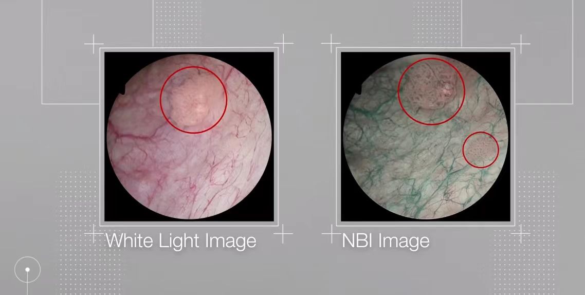
Narrowband imaging is a revolutionary optical image enhancement technology whose core idea is "selective seeing." It moves away from white light, which provides a continuous spectrum, and instead takes advantage of specific physical properties. ISP will instruct the light source system to emit only two specific narrowband wavelengths of light: one is blue light around 415nm, and the other is green light around 540nm. According to the characteristics of biological tissue, shorter blue light can only penetrate the surface layer of mucous membranes, so it will be largely absorbed by the hemoglobin in the capillary network on the surface, making it appear dark (usually tan). Green light with a slightly longer wavelength can penetrate into the submucosa, be absorbed by deeper blood vessels, and appear in different colors (usually cyan).
Key roles of ISP:
The implementation of NBI completely relies on the precise control and high-speed operation of ISP.
- Light source synchronization: ISP must be perfectly synchronized with the light source controller to drive the LED to perform ultra-high-speed stroboscopic switching between two wavelengths.
- Frame sequence processing: At each switching moment, the ISP synchronously instructs the image sensor to capture, thereby obtaining a sequence of monochromatic image frames illuminated only by blue light or green light.
- Image synthesis and pseudo-color rendering: After ISP receives these monochrome frames, it does not simply superimpose them, but analyzes and enhances them, and performs pseudo-color rendering based on the preset medical model - rendering the superficial blood vessels under blue light into tan, and rendering the deep blood vessels under green light into cyan, and finally synthesizes a real-time image that can clearly show the "two-layer structure" of mucosal blood vessels.
Clinical significance:
In precancerous and early cancerous tissues, the superficial blood vessels will undergo abnormal changes such as proliferation, distortion, and expansion. Under traditional white light, these changes are often difficult to detect due to mucosal color interference. By allowing these abnormal blood vessel networks to "surface" clearly, NBI technology has greatly improved the detection rate of early cancers in the digestive tract, respiratory tract and other parts of the body, and is a veritable "lighthouse" for early cancer screening.
Technical advantages:
Through innovative algorithms, we have achieved faster image processing speed and lower power consumption, while significantly improving the contrast and clarity of early lesions, providing doctors with a more reliable basis for diagnosis.
2. Blood vessels and color enhancement
How it works:
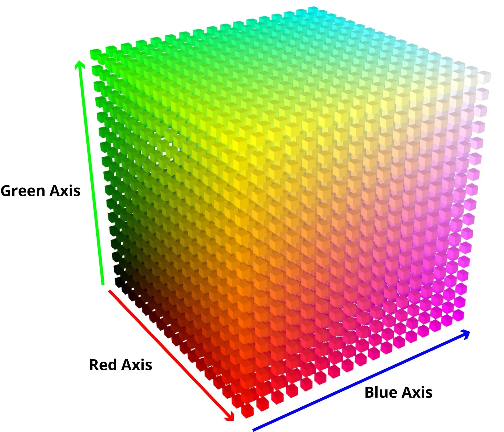
Different from the special mode of NBI, the blood vessel and color enhancement algorithm works on regular white light images, aiming to optimize its visual performance and highlight the blood vessel structure. Its core lies in the refined operation of color space. ISP will convert the RGB (red, green, and blue) images collected by the camera into a color space (such as HSV/HSL) that is more in line with human eye perception in real time, and then make non-linear adjustments to the hue (Hue) representing "color" and the saturation (Saturation) representing "vividness".
Key roles of ISP:
The ISP will execute a 3D color look-up table (3D-LUT), which is a "color secret book" calibrated in advance by image engineers and medical experts. When the ISP identifies a specific range of reds that belong to blood, it queries the 3D-LUT to make those reds more saturated and purer while suppressing the color of the surrounding tissue, allowing the network of blood vessels to stand out as clearly as a river on a map. The entire process is completed by hardware in the pipeline to ensure real-time performance.
Clinical significance:
This function is crucial for judging inflammation (usually accompanied by blood vessel congestion and dilation), finding tiny bleeding points, assessing the blood supply of tumors, and assisting in hemostasis during surgery.
Technical advantages:
Our blood vessel and color enhancement algorithm uses a unique color space mapping model and adaptive enhancement technology. Compared with traditional algorithms, it can more accurately highlight the blood vessel network at all levels without introducing color distortion, and the detail display capability is improved by more than 30%, greatly improving the recognition rate of tiny blood vessels.
3. Dynamic contrast enhancement
How it works:
The surface of the human body's internal cavity is moist and has a rugged structure, which can easily form dazzling high-light reflections and deep tissue fold shadows under the lens. This results in an image with an extremely large dynamic range (the ratio of lightest to darkest). In order to see details in highlights and shadows simultaneously, ISP uses high dynamic range (HDR) synthesis and local tone mapping (Local Tone Mapping) technology.
Key roles of ISP:
- Multiple exposure synthesis: ISP can control the sensor to continuously take multiple (usually 2-3) photos with different exposure times in a very short time: a short exposure captures the details of the highlight area, and a long exposure captures the details of the shadow area. The ISP then intelligently fuses these photos into one raw image containing ultra-high dynamic range.
- Intelligent Tone Mapping: Due to the limited dynamic range of displays, ISPs also need to perform a critical "tone mapping" step to compress HDR data into the display range. Advanced ISP will use local tone mapping, which divides the image into multiple areas, independently analyzes the brightness distribution of each area, and adjusts the contrast adaptively. This can suppress highlights and brighten dark areas, while retaining texture details in the respective areas, allowing doctors to obtain a clear view of "what you see is everything".
Clinical significance:
This function ensures that doctors will not miss early lesions under high-light areas due to strong reflection, nor ignore abnormalities hidden deep in folds due to shadows, greatly improving the comprehensiveness and accuracy of examinations.
Technical advantages:
We use edge-preserving contrast enhancement technology combined with multi-exposure fusion to achieve a dynamic range of over 100dB. Even in extreme scenes where high-light reflections and deep wrinkles coexist, we can ensure that every detail is clearly visible and no detail is missed.
4. Noise reduction and sharpening
How it works:
This is the art of balancing spear and shield. Noise reduction aims to remove image noise produced in low light, while sharpening (edge enhancement) aims to make images look clearer. However, excessive noise reduction will erase details and create a "smeared feeling"; excessive sharpening will produce an unnatural "halo" and amplify noise.
Key roles of ISP:
Modern medical ISP uses an adaptive hybrid noise reduction and sharpening strategy.
- 3D noise reduction: ISP will simultaneously analyze the spatial dimension (Spatial) of a single frame image and the temporal dimension (Temporal) of consecutive multi-frame images. It determines that if a pixel is similar to its surroundings in space (such as a flat mucosal area) and jumps randomly in time, then it is likely to be noise and should be smoothed.
- Content adaptive sharpening: ISP will first analyze the image content to identify which are real tissue edges, which are flat areas, and which are noise. Then, it will "smartly" selectively sharpen only the identified tissue edges, while enhancing noise reduction in flat areas, thereby minimizing artifacts and unnaturalness while improving clarity.
Clinical significance:
An excellent noise reduction and sharpening system can provide doctors with an image that is both pure and clear while retaining the original texture of the tissue. This is crucial for observing the fine texture of the mucosa and judging the clarity of the lesion boundary.
Technical advantages:
The "spatial and temporal domain joint noise reduction" and "content adaptive sharpening" algorithms we developed can intelligently identify noise and tissue texture in images. While effectively removing more than 95% of the noise, the sharpening of tissue edges is "just right", perfectly avoiding the "halo" and "artifact" problems caused by traditional sharpening technology, and achieving the best balance between image purity and the true texture of the tissue.
Chapter 3: Driver Core: Strategic Choice of ISP Hardware Platform
If exquisite algorithms are the “soul” of endoscope ISP, then the hardware platform that hosts these algorithms is its “body”. The choice of hardware not only determines the ceiling of ISP performance, but is also a strategic decision related to product positioning, R&D cycle, cost control and even future upgrade potential. In this decision, the industry faces, for example, a trade-off between "mass-produced sports car engines" and "customized F1 racing engines."
1. SoC platform: mature and efficient integrated solution
SoC (System-on-Chip) is like the highly integrated engine that powers mainstream sports cars. It packages the market-proven CPU core, dedicated ISP image processing pipeline, memory controller and rich input and output interfaces on a small silicon chip.
Typical choice for domestic market
In the domestic endoscope market, a significant trend is that many manufacturers cleverly "cross-border" utilize domestic SoC chips that have been proven on a large scale in other mature fields (such as high-end security, TV boxes, artificial intelligence Internet of Things). This not only accelerates product iteration, but also greatly improves the cost-effectiveness of the solution.
- Rockchip: Known for its powerful multimedia processing capabilities and general computing performance. For example, flagship chips such as the classic RK3399 and the new generation of integrated NPU (neural network processor) RK3568 and RK3588 are widely used in endoscope host systems that need to run complex operating systems (such as Android/Linux), support high-definition display and rich human-computer interaction. Their powerful CPU/GPU/NPU clusters allow them to not only process images, but also easily host upper-layer applications such as AI-assisted diagnosis.
- Hisilicon: Originating from the world's top security surveillance (IPC) field, Hisilicon's SoCs (such as the Hi35xx series) are highly favored for their internally integrated high-quality, highly optimized ISP pipeline. These ISPs are designed to adapt to all-weather, low-light, and highly dynamic security scenarios, and their technical features coincide with the needs of endoscopes. Choosing the HiSilicon solution means, to a certain extent, that you directly get a set of "out-of-the-box" high-performance imaging core. The success of this "cross-border" strategy lies in the fact that it cleverly leverages the huge R&D investment and large-scale cost advantages of consumer/industrial-grade chips, allowing medical equipment manufacturers to focus more on clinical needs, software applications and the polishing of optical systems.
Core advantages: standardization and high efficiency
For developers, choosing SoC means getting a "turnkey" solution. The hardware design has been greatly simplified, and the focus of research and development has returned to the more familiar software level. Through the SDK (software development kit) provided by the manufacturer, engineers use high-level languages such as C/C++ to call and configure the ISP module solidified inside the SoC, just like filling in a menu, checking the required functions and fine-tuning parameters. This model greatly shortens the product launch cycle, and when shipments reach hundreds of thousands or even millions, the cost advantage brought by its scale effect is unparalleled.
Scenario application: portable and disposable main force
These characteristics of SoC make it the first choice for portable/handheld endoscopes, capsule endoscopes, and extremely cost-sensitive disposable endoscopes. In these applications, SoC is sufficient to provide high-quality high-definition images, and its low-power consumption characteristics also perfectly meet the needs of mobility and miniaturization.
Inherent limitations: the “shackles” to flexibility
However, SoC's "menu-style" development model also means that its flexibility is limited. The core algorithms of ISP, such as noise reduction, wide dynamic range, etc., are "hard-coded" in the hardware logic. Developers can adjust the "strength" of the algorithm, but they cannot change the algorithm itself. If a revolutionary new algorithm comes out, SoC-based devices will not be able to obtain it through software upgrades and will have to wait for the release of the next generation chip. This is undoubtedly a "shackle" for high-end equipment that pursues cutting-edge technology.
2. FPGA platform: the “blank canvas” for the pursuit of perfection and the future
Completely opposite to SoC, FPGA (Field-Programmable Gate Array) is a "blank canvas" on which creativity can be unleashed. It is a customized engine born for F1 racing cars that pursues ultimate performance. It is composed of a large number of reprogrammable logic units. Developers are no longer "software engineers" but become "chip designers".
Mainstream FPGA choices on the market
In the field of high-end medical equipment with extremely high performance and reliability requirements, the FPGA market is mainly dominated by two international giants: Xilinx, which has been acquired by AMD, and Altera, which is affiliated with Intel. When domestic manufacturers choose FPGA solutions, they mostly focus on the product series of these two companies.
- Xilinx (AMD) solution: With its strong ecosystem and rich product line, Xilinx occupies a leading position in the field of high-end medical imaging.
- Kintex series: such as Kintex-7 and Kintex UltraScale series, are "frequent users" of mid-to-high-end endoscope hosts. They achieve an excellent balance between performance, power consumption and cost, and are sufficient to support complex 4K ISP pipelines and AI algorithms.
- Zynq series: especially Zynq UltraScale+ MPSoC, has become the star solution for the new generation of high-end hosts. It integrates a quad-core ARM Cortex-A53 processor and powerful FPGA logic on a single chip, enabling the "hybrid" mode we mentioned earlier. The CPU part can run Linux or Android systems smoothly and is responsible for user interaction and network functions; while the FPGA part is dedicated to real-time, low-latency transmission and processing of multi-channel videos such as 4K/8K, 3D, and fluorescence.
- Intel (Altera) solution: As another giant, Intel's FPGA also provides a very competitive choice.
- Arria series: Directly benchmarking Xilinx’s Kintex series, it provides another high-performance option for mid-to-high-end systems.
- Cyclone V SoC: Similar to Xilinx's Zynq-7000 series, it integrates a dual-core ARM Cortex-A9 processor and FPGA logic, providing a mature and reliable platform for mid-range devices that require both system management and hardware acceleration.
- Domestic FPGA solutions: In recent years, domestic FPGA manufacturers represented by Pango and Gowin have developed rapidly and have achieved remarkable results in the medium and low-density consumer and industrial markets. However, in high-end medical applications such as endoscope hosts, which have extremely strict requirements on logic scale, processing performance, high-speed interfaces and long-term reliability, domestic FPGAs still lag behind the two international giants in terms of performance and ecological maturity. Currently, they are mainly used for some peripheral auxiliary functions or relatively simple entry-level equipment. Breaking through high-end medical applications is a key fortress that domestic FPGA manufacturers need to overcome in the future.
Core advantages: Customization and ultra-high performance
Using hardware description languages such as Verilog or VHDL, developers can build a unique ISP processing pipeline directly on the FPGA that is completely customized for their own algorithms. This hardware-level parallel architecture enables dozens of processing steps to work together in the same clock cycle, achieving ultra-high throughput and microsecond-level deterministic latency that cannot be achieved by software. This is crucial for top surgical systems that need to handle multiple video streams such as 4K/8K, 3D, and fluorescence at the same time.
Scenario application: the best choice for high-end flagships
The ultimate performance and unparalleled flexibility of FPGA make it an inevitable choice for high-end multi-functional surgical endoscope hosts, cutting-edge scientific research platforms, and flagship products of various brands. Enterprises can implement their own unique patented algorithms as technical barriers on FPGA, thereby standing out in the fierce market competition. More importantly, the reprogrammable nature of FPGA means that the device is "future-proof" and can continue to bring new features and better image quality to users through firmware upgrades even after it is sold.
High price: the threshold of technology and cost
The cost of this “blank canvas” is high. The development of FPGA not only requires a long cycle and a professional hardware design team, but the cost of the chip itself and supporting development software is also much higher than that of SoC. In addition, its powerful performance is often accompanied by higher power consumption and cooling requirements.
3. The Way of Integration: The Rise of the “Hybrid Power” Model
In reality, more and more top-end devices are beginning to adopt a "hybrid" model, which uses a heterogeneous SoC chip that integrates a CPU core (ARM) and FPGA programmable logic. This integration solution achieves the best of both worlds: non-real-time tasks such as complex operating systems, user interfaces, and network communications are handled by more suitable CPU cores, adopting an efficient software development model; while the ISP image processing pipeline, which has extremely demanding requirements on latency and throughput, is implemented in the FPGA logic part, giving full play to its hardware parallel processing advantages. This not only ensures the ultimate performance of the system, but also takes into account the flexibility and efficiency of development, and is becoming the mainstream development direction of the next generation of high-end endoscope platforms.
All in all, the choice of hardware platform is not a simple technical team, but a comprehensive strategy based on multi-dimensional considerations such as product market positioning, performance goals, cost budget and R&D strength. From efficient and economical SoCs, to ultimate FPGAs, to eclectic hybrid architectures, this path of hardware evolution clearly reflects the firm pace of endoscope technology developing toward more precision, intelligence, and diversification.
Chapter 4: China’s Power: Development Status and Challenges of Local Endoscopy ISPs
In the past, the global high-end endoscope market has long been dominated by several traditional Japanese and German giants, and their core technologies, especially ISP and image sensors (CMOS), have formed deep barriers. However, in recent years, driven by strong market demand and national policies, China's endoscope industry chain is accelerating its rise.
Development status:
- The wave of “domestic substitution” is accelerating: As the country attaches great importance to the independent control of high-end medical equipment, the entire industry chain, from complete machine manufacturers to upstream chip design companies, is actively promoting the localization of core components. Local ISP solutions are gradually entering the market and beginning to replace imported solutions in some mid- to low-end and even mid-to-high-end equipment.
- Technical level is catching up quickly: Domestic leading medical equipment companies are no longer satisfied with producing basic high-definition products, and are making every effort to move towards high-end fields such as 4K and fluorescence imaging. Behind this, it is inseparable from the rapid iteration and progress of local ISP chip design companies in algorithms and chip capabilities.
- The synergy effect of the industrial chain is beginning to show: The cooperation between complete machine manufacturers, ISP solution providers, and CMOS sensor manufacturers is becoming increasingly close, forming a good industrial ecology. The complete machine manufacturer provides valuable clinical needs and image debugging experience, while the chip company provides a powerful computing platform and algorithm support to jointly polish the product.
Challenges faced:
- There is still a gap in core technology: despite significant progress, in terms of the "Know-How" of image tuning (that is, how to adjust image effects to the best state based on a large amount of clinical feedback), domestic companies still need time to settle compared with international giants who have accumulated decades of experience.
- Dependence on key upstream components: In fields such as the top CMOS image sensors, the problem of external dependence still exists, which to a certain extent limits the performance upper limit of the most high-end domestic endoscopes.
- Brand and market trust: The medical field has extremely high requirements for product stability and brand trust. To establish the same level of trust as international giants in top hospitals, domestic brands need long-term market testing and reputation accumulation.
Chapter 5: Future Trend: Smarter and more popular “Chinese Eyes”
Looking to the future, endoscopic ISP technology is evolving towards intelligence, precision and popularization, which is also a key opportunity for domestic enterprises to achieve "overtaking in corners".
- Deep integration of AI and ISP: This is industry consensus. Future ISPs will have built-in AI units that can not only realize intelligent optimization of image parameters, but also run auxiliary diagnostic algorithms to prompt doctors to pay attention to suspicious lesions in real time, becoming a true "intelligent vision system."
- Moving towards higher image quality: From 4K to 8K, from 2D to 3D stereoscopic vision, higher-quality images can provide doctors with unprecedented details and depth perception, especially helping to improve the accuracy of surgical operations.
- Portability and integration: With the advancement of technology, powerful ISPs will be integrated into smaller portable devices, allowing endoscopy to go out of the examination room and be used in more scenarios such as bedside, emergency, primary care, etc.
- The rise of disposable endoscopes: To address the risk of cross-infection and reduce maintenance costs, the market for disposable endoscopes is growing rapidly. This will generate huge demand for ultra-small, low-cost, high-performance SoC ISP, which is an important opportunity for local chip companies.